
Amiodarone Pulmonary Toxicity Risk Calculator
Patient Risk Assessment Tool
This calculator helps determine the risk of amiodarone pulmonary toxicity based on key clinical factors and treatment parameters. Higher risk indicates need for more intensive monitoring.
Key Takeaways
- Amiodarone can cause lung injury ranging from mild cough to life‑threatening fibrosis.
- Risk rises with daily doses >400 mg, treatment longer than 6 months, and patient age >60.
- Early clues: new dyspnea, non‑productive cough, low‑grade fever, or subtle chest‑X‑ray changes.
- Confirming toxicity relies on high‑resolution CT, pulmonary function tests, and ruling out infection.
- Management = stop drug, consider corticosteroids, and monitor lung function every 3 months for the first year.
When a clinician prescribes Amiodarone is a class III anti‑arrhythmic medication used for rhythm control in Atrial fibrillation and Ventricular tachycardia. While it’s highly effective, its long half‑life (≈50 days) and iodine‑rich structure make the lungs a frequent collateral target. This article walks through the anatomy of amiodarone pulmonary toxicity, how to spot it early, and steps to keep patients breathing easy.
What is Pulmonary Toxicity?
Pulmonary toxicity refers to any drug‑induced injury to lung tissue that impairs gas exchange. With amiodarone, the most common patterns are Interstitial pneumonitis and, in chronic cases, Pulmonary fibrosis. Both can mimic infection, heart failure, or other interstitial lung diseases, which is why a systematic approach matters.
Who Is at Highest Risk?
- Daily dose ≥ 400 mg (the risk curve steepens around 600 mg).
- Treatment duration > 6 months.
- Age > 60 years.
- Pre‑existing lung disease (COPD, sarcoidosis).
- Concomitant high‑oxygen therapy or radiation.
- Renal or hepatic impairment that slows drug clearance.
Patients with these factors should have baseline lung evaluation before starting therapy.
Early Signs & Symptoms to Watch For
Symptoms often develop insidiously, so a high index of suspicion is key. Typical presentations include:
- Progressive dyspnea on exertion (often the first clue).
- Dry, non‑productive cough.
- Low‑grade fever without obvious source.
- Chest discomfort that worsens with deep breathing.
- Unexplained fatigue or weight loss.
If any of these appear after the first few months of therapy, pause and order imaging.
Diagnostic Work‑up
The goal is to confirm drug‑related injury while excluding infection, heart failure, or other interstitial lung diseases.
- Chest X‑ray: May show diffuse interstitial infiltrates, but can be normal in early disease.
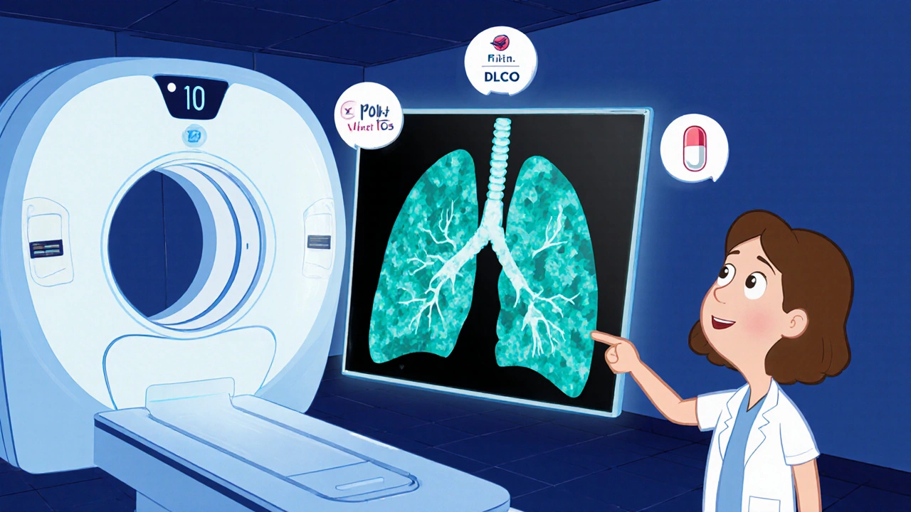
- High‑resolution computed tomography (HRCT): High-resolution computed tomography (HRCT) reveals ground‑glass opacities, crazy‑paving, or early fibrosis-patterns highly suggestive of amiodarone injury.
- Pulmonary function tests (PFTs): Pulmonary function test (PFT) typically shows a restrictive pattern with a reduced diffusing capacity for carbon monoxide (DLCO).
- Bronchoalveolar lavage (BAL) or lung biopsy: Reserved for ambiguous cases; BAL may show foamy macrophages, while biopsy confirms interstitial pneumonitis or fibrosis.
- Serum amiodarone level: Not routinely diagnostic, but a level > 2.5 µg/mL often correlates with toxicity.
Management Strategies
Prompt action can reverse most early changes.
- Discontinue amiodarone: Switch to an alternative anti‑arrhythmic (e.g., dofetilide, sotalol) if rhythm control remains necessary.
- Corticosteroid therapy: For moderate‑to‑severe pneumonitis, start prednisone 0.5‑1 mg/kg/day, taper over 4‑6 weeks based on clinical response and imaging.
- Supportive care: Supplemental oxygen, pulmonary rehabilitation, and monitoring for secondary infections.
- Follow‑up monitoring: Repeat HRCT and PFTs at 4‑6 weeks, then every 3 months for the first year.
In cases where fibrosis has set in, steroids are less effective; focus shifts to symptom control and lung transplantation evaluation if appropriate.
Prevention & Monitoring Checklist
| Parameter | Recommended Frequency | Typical Threshold |
|---|---|---|
| Baseline chest X‑ray | Before starting therapy | Normal |
| HRCT | 6 months, then annually if dose > 400 mg | No new ground‑glass opacities |
| PFT (including DLCO) | Every 3 months for the first year | DLCO ↓ > 15 % from baseline |
| Serum amiodarone level | Every 6 months if dose > 400 mg | ≤ 2.5 µg/mL |
| Clinical symptom screen | At each cardiology visit | Any new dyspnea or cough |

When to Involve a Specialist
If the patient exhibits any of the following, refer to a pulmonologist without delay:
- Rapidly worsening dyspnea.
- Oxygen saturation < 90 % on room air.
- Radiographic progression despite drug withdrawal.
- Unclear diagnosis after initial work‑up.
Case Vignette
Mr. J., 68, started amiodarone 600 mg/day for recurrent ventricular tachycardia. After 8 months he noted a “tightness” in his chest and a dry cough. A routine chest X‑ray was subtle; HRCT revealed diffuse ground‑glass opacities. PFTs showed a 20 % drop in DLCO. The cardiology team stopped amiodarone, started prednisone 40 mg daily, and switched rhythm control to catheter ablation. Six weeks later, his symptoms resolved, and repeat HRCT showed marked improvement. This illustrates the value of early imaging and swift drug discontinuation.
Bottom Line
Amiodarone remains a cornerstone for life‑threatening arrhythmias, but its pulmonary toxicity demands vigilance. By screening high‑risk patients, recognizing the subtle early signs, and acting decisively-withdrawal plus, when needed, corticosteroids-clinicians can prevent irreversible lung damage.
What dose of amiodarone is considered high risk for lung toxicity?
Daily doses of 400 mg or higher, especially when used for more than six months, markedly increase the chance of pulmonary injury.
Can pulmonary toxicity develop after stopping amiodarone?
Yes. Because the drug’s half‑life is long, lung damage can continue to evolve for weeks after discontinuation. Ongoing monitoring is essential.
Is a chest X‑ray enough to rule out amiodarone‑induced lung disease?
No. Early toxicity often shows normal X‑ray findings. High‑resolution CT is far more sensitive and should be used when suspicion exists.
Should I always switch to another anti‑arrhythmic if toxicity occurs?
If rhythm control is still needed, an alternative such as sotalol, dofetilide, or catheter ablation should be considered. The choice depends on the underlying arrhythmia and patient comorbidities.
How long does steroid therapy usually last for amiodarone‑induced pneumonitis?
A typical course is 4‑6 weeks of tapering, guided by symptom resolution and imaging improvement. Some patients may need a slower taper.

Gotta get a baseline chest X‑ray before you even start amiodarone, it makes later comparisons way easier.
Even if the patient feels fine, the imaging gives you a reference point.
Age and dose matter, so a good initial picture can catch subtle changes early.
And hey, a quick PFT at baseline isn’t a hassle – it pays off if toxicity shows up later.
Just keep the paperwork tidy, it’ll save you a lot of headaches down the road.
I've seen a few folks skip that initial imaging and then scramble when a dry cough shows up months later.
Getting that baseline is a simple habit that pays dividends.
It's especially critical for patients over 60 or those on higher doses.
Having the early picture also guides the decision on when to order a high‑resolution CT.
When you think about amiodarone pulmonary toxicity, the first thing that comes to mind is the insidious nature of its presentation.
Patients often report a mild, non‑productive cough that they brush off as a cold, yet underneath lies a cascade of inflammatory changes.
Because the drug has a half‑life of about fifty days, the toxic effects can linger even after the medication is stopped, making timely recognition essential.
Baseline chest X‑ray and pulmonary function tests provide a reference point that clinicians can compare against future studies, and this practice cannot be overstated.
High‑resolution CT, on the other hand, is far more sensitive; ground‑glass opacities or crazy‑paving patterns are hallmarks that should raise suspicion immediately.
In my experience, ordering a CT when DLCO drops more than fifteen percent from baseline often catches the disease before fibrosis sets in.
Bronchoalveolar lavage may reveal foamy macrophages, which, while not pathognomonic, support the diagnosis when other causes have been excluded.
It is also vital to rule out infection, heart failure, and other interstitial lung diseases because the treatment pathways diverge significantly.
Once toxicity is confirmed, the cornerstone of management is discontinuation of amiodarone, followed by a judicious taper of prednisone, typically starting at half to one milligram per kilogram per day.
The steroid dose should be tailored to the severity of the pneumonitis, and patients must be monitored closely for side effects like hyperglycemia or mood swings.
Supportive care, including supplemental oxygen and pulmonary rehabilitation, can improve quality of life while the lungs heal.
Follow‑up imaging at four to six weeks, and then every three months for the first year, gives a clear picture of recovery or progression.
If fibrotic changes become established, corticosteroids lose their efficacy, and the focus shifts toward symptom management and, in severe cases, evaluation for lung transplantation.
Education of patients is equally important; they should be instructed to report any new dyspnea, cough, or fever promptly, even if symptoms seem mild.
In summary, a systematic approach-baseline testing, vigilant monitoring, early imaging, and rapid drug withdrawal-provides the best chance to prevent irreversible lung damage.
Only seasoned pulmonologists truly grasp the nuance of amiodarone‑induced interstitial changes, the rest are just guessing at generic coughs. The diagnostic algorithm outlined here is elementary, yet many still rely on plain chest X‑rays, missing the high‑resolution detail that reveals the pathology. If you want to avoid the inevitable pitfalls, adopt a rigorous monitoring schedule from day one-no shortcuts. 🌟
i think its smart to keep an eye on DLCO early on, it can warn before big problems.
Implementing a structured follow‑up protocol aligns with evidence‑based practice and minimizes unnecessary hospitalizations.
Providing patients with clear guidance on symptom monitoring empowers them to seek care promptly, which can dramatically improve outcomes.
Totally agree, a quick reminder at each cardiology visit about new cough or breathlessness can make a big difference.
Amiodarone can hurt the lungs if you take too much for too long.
The way the drug stealthily accumulates iodine in alveolar walls feels almost theatrical, a silent antagonist lurking beneath the surface.
One moment the patient is fine, the next they gasp for air and wonder why.
This slow‑burn toxicity isn't just a footnote; it's a full‑blown drama that demands early detection.
High‑resolution CT is the spotlight that reveals the hidden plot, while routine X‑rays play the understudy.
When the curtain finally rises on fibrosis, the options narrow dramatically, making prevention the star of the show.
While the narrative emphasizes early imaging, one could argue that excessive CT scans expose patients to unnecessary radiation, suggesting a more measured approach based on clinical symptomatology.
Sounds exaggerated.
Let's keep the conversation lively-remember, early detection, patient education, and a solid monitoring plan are the three pillars that keep amiodarone‑related lung injury at bay! 🚀